
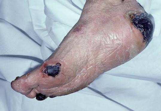
Eschar on wound refers to a thick layer of dead (necrotic) or devitalized tissue that develops over or within an injured area. Dealing with skin injuries, pressure sores, or chronic ulcers can be challenging, especially when you notice a dark, hard, or leathery layer forming over the affected area.
While many people mistake this layer for a standard scab, it is a distinctly different medical condition. Understanding what an eschar on wound is, why it forms, how clinicians manage it, and when to seek urgent care can help patients and caregivers make safe decisions and avoid severe health complications.
What Is Eschar on Wound?
Eschar on wound is composed of dead skin cells, collapsed collagen fibers, proteins, and dried inflammatory fluid. Unlike healthy tissue, necrotic tissue has completely lost its vascular supply, meaning oxygen and essential nutrients can no longer reach the cells. Consequently, this tissue dies off and forms a firm, often dark barrier over the wound bed.
                    ┌─────────────────────────┐
                    │      Wound Bed          │
                    └────────────┬────────────┘
                                 │
                    ┌────────────▼────────────┐
                    │ Loss of Vascular Supply │
                    └────────────┬────────────┘
                                 │
                    ┌────────────▼────────────┐
                    │ Cellular Necrosis/Death │
                    └────────────┬────────────┘
                                 │
                    ┌────────────▼────────────┐
                    │  Eschar on Wound Forms  │
                    └─────────────────────────┘
Eschar vs. Scab: Key Differences
It is common to confuse an eschar on wound with a simple scab, but their composition, depth, and clinical implications are entirely different:
| Feature | Eschar on Wound | Standard Scab |
| Composition | Dead, necrotic body tissue and structural proteins | Dried blood, serum, and cellular debris |
| Depth | Involves deeper skin layers, subcutaneous fat, or muscle | Primarily superficial (epidermis/upper dermis) |
| Appearance | Black, dark brown, or tan; thick, leathery, or crusty | Reddish-brown or dark brown; thin and brittle |
| Primary Causes | Deep burns, pressure ulcers, diabetic foot ulcers, severe trauma | Minor cuts, scrapes, abrasions, superficial wounds |
| Management | Often requires professional evaluation or debridement | Sheds naturally as underlying skin heals |
What Does an Eschar on Wound Look Like?
The physical characteristics of an eschar on wound vary depending on the wound type, moisture levels, and the age of the necrotic tissue. Common physical features include:
Color: Typically dark brown, black, tan, or deep grey.
Texture: Often dry, hard, firm, and leathery. In some cases, if fluid builds up beneath it, it can become soft, wet, or boggy.
Attachment: Firmly adhered to the underlying wound bed and surrounding healthy tissue.
Sensation: Lacks sensation directly over the dead tissue because local nerve endings are destroyed or covered. However, the surrounding healthy skin may be tender or painful.
The Problem of Hidden Depth
One of the biggest challenges an eschar on wound presents to medical providers is that it acts as a mask. Because it forms a thick, opaque cap over the injury, clinicians cannot inspect the underlying tissue directly.
In pressure injuries (bedsores), a wound completely covered by an eschar on wound is classified as Unstageable. Until a portion of the dead tissue is softened or removed, it is impossible to determine whether the tissue damage extends into fat, muscle, or bone.
What Causes an Eschar on Wound?
An eschar on wound develops whenever localized blood flow is severely restricted or tissue is destroyed faster than the body can repair it. The primary causes include:
1. Pressure Injuries (Bedsores)
Individuals with limited mobility—such as bedridden patients or wheelchair users—are at high risk for pressure injuries. Sustained pressure over bony prominences reduces blood supply, leading to ischemic tissue death. Common sites include:
- Heels and ankles
- Sacrum (tailbone) and hips
- Elbows and shoulder blades
- Back of the head
2. Diabetic Foot Ulcers
Diabetes can cause nerve damage (neuropathy) and peripheral artery disease (PAD). Reduced blood flow paired with diminished pain sensation means minor foot injuries often go unnoticed, progressing to deep ulcers with an eschar on wound.
3. Burns
Full-thickness (third-degree) and deep partial-thickness (second-degree) burns destroy the epidermis and dermis, charring proteins and cellular structures to form a thick eschar on wound.
Critical Note: In severe circumferential burns (burns wrapping entirely around a limb or the torso), shrinking tissue can act like a tight band, constricting blood vessels or hampering breathing. This medical emergency requires an urgent surgical release known as an escharotomy.
4. Severe Trauma and Vascular Disease
Crush injuries, surgical wounds with poor edge healing, or advanced Peripheral Arterial Disease (PAD) impair local circulation, causing skin and sub-dermal tissues to die and turn into an eschar on wound.
How Is an Eschar on Wound Treated?
There is no single approach to managing an eschar on wound. Treatment depends on blood circulation, the presence of infection, wound location, and overall patient health.
The Rule of Unstable vs. Stable Eschar
Stable, Dry Eschar (e.g., on an Ischemic Heel): If an eschar on wound located on a heel or lower extremity is dry, firmly adhered, without redness (erythema), swelling, or drainage, clinicians often leave it intact. In patients with poor blood circulation, the dry tissue serves as a natural, sterile biological cover. Removing it prematurely without adequate blood flow can create an open wound that cannot heal, increasing infection risk.
Unstable, Wet, or Infected Eschar: If the eschar on wound is soft, boggy, draining fluid, foul-smelling, or surrounded by inflamed skin, it must be addressed promptly. Soft, wet necrotic tissue breeds bacteria and must be removed through debridement.
Methods of Medical Debridement
Debridement is the medical process of removing non-viable tissue to expose healthy, healing tissue underneath.
Why You Must Never Remove an Eschar on Wound Yourself
Warning: Never attempt to pick, cut, scrape, or peel an eschar on wound at home using scissors, tweezers, or blades.
Attempting self-debridement carries major health risks:
- Severe Hemorrhage: Unintentional cutting into underlying viable blood vessels can cause heavy bleeding.
- Deep Tissue Damage: Without professional training, healthy muscle, fat, or tendons can be damaged.
- Severe Infection: Non-sterile home tools introduce dangerous bacteria deep into open tissue layers.
Warning Signs: When to Seek Immediate Medical Care
While some chronic wounds heal slowly under monitoring, certain warning signs indicate a dangerous infection or tissue breakdown. Seek urgent medical care if you observe:
- Spreading Redness or Heat: Expanding warmth or red streaks moving away from the wound edge.
- Purulent or Foul-Smelling Drainage: Green, yellow, or cloudy fluid leaking from beneath the eschar on wound, especially with a foul odor.
- Systemic Symptoms: Fever, chills, dizziness, confusion, or nausea.
- Increasing Severe Pain: Sudden or worsening pain surrounding the wound site.
- Boggy or Soft Texture: A dry eschar on wound suddenly becoming soft, mushy, or elevated.
Leave a Reply