
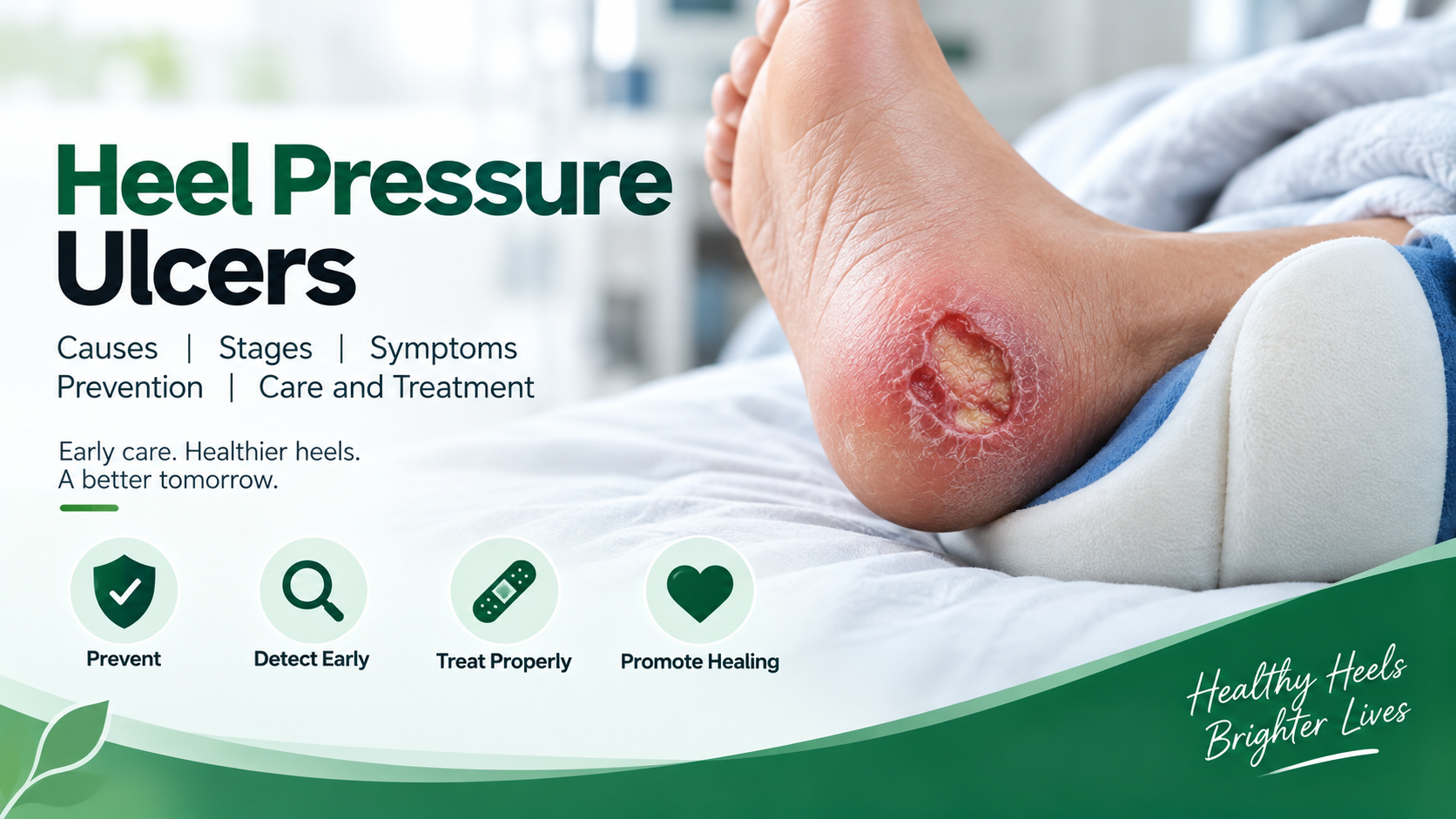
Heel pressure ulcers, also called heel pressure injuries or heel bedsores, are wounds that develop when prolonged pressure on the heel reduces blood flow and damages the skin and underlying tissues. They are particularly common in people who are bedridden, have limited mobility, use wheelchairs, or have poor circulation.
Because the heel has relatively little soft tissue between the skin and the underlying bone, it can be especially vulnerable to pressure-related damage. Early identification and proper pressure relief can help prevent a minor skin change from developing into a deeper wound.
What Are Heel Pressure Ulcers?
A heel pressure ulcer is an area of skin and tissue damage that develops when continuous or repeated pressure is placed on the heel.
When pressure remains on the heel for an extended period, blood circulation to the tissues may become reduced. This limits the supply of oxygen and nutrients and can eventually cause tissue damage.
Heel pressure injuries may initially appear as a change in skin color, temperature, firmness, or tenderness. In some cases, significant tissue damage can develop beneath the skin before an obvious open wound becomes visible.
The heels are considered an important pressure point because they are a prominent bony area with relatively limited cushioning.
What Causes Heel Pressure Ulcers?
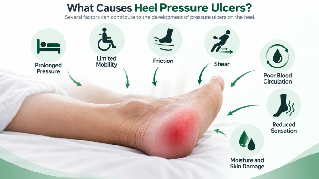
Common factors that can contribute to the development of heel pressure ulcers.
1. Prolonged Pressure
The primary cause is prolonged pressure on the heel.
A person who remains in the same position in bed for a long time may continuously place pressure on the back of the heel. If the pressure is not relieved, blood flow can become compromised and tissue damage may develop.
2. Limited Mobility
People who cannot move or reposition themselves independently are at greater risk.
This includes people who are:
- Bedridden
- Recovering from surgery
- Living with mobility limitations
- Using a wheelchair for extended periods
- Experiencing weakness after serious illness
3. Friction
Repeated rubbing between the heel and bedding, clothing, or other surfaces can damage the outer layer of skin.
4. Shear
Shear can occur when the body moves in one direction while the skin remains relatively fixed against the surface.
For example, sliding down in bed can place additional stress on tissues.
5. Poor Blood Circulation
Reduced circulation can make the heel more vulnerable to pressure-related damage and may also slow healing once an injury develops. People with peripheral vascular disease may have increased risk of pressure ulcers affecting the feet and heels.
6. Reduced Sensation
People who have reduced sensation may not notice discomfort or early skin changes and therefore may remain under pressure for longer periods.
7. Moisture and Skin Damage
Excessive moisture can weaken the skin and make it more susceptible to breakdown, particularly when combined with pressure and friction.
Who Is at Risk of Heel Pressure Ulcers?
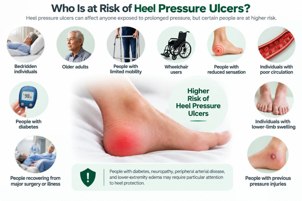
Heel pressure ulcers can affect anyone exposed to prolonged pressure, but the risk may be higher among:
Visual guide to the different stages and classifications of heel pressure injuries.
- Bedridden individuals
- Older adults
- People with limited mobility
- Wheelchair users
- People with reduced sensation
- Individuals with poor circulation
- People with diabetes
- Individuals with lower-limb swelling
- People recovering from major surgery or illness
- Individuals who cannot reposition themselves independently
- People with previous pressure injuries
People with diabetes, neuropathy, peripheral arterial disease, and lower-extremity edema may require particular attention to heel protection.
Common Signs and Symptoms of Heel Pressure Ulcers
Early identification is important because pressure injuries can worsen if pressure continues.
Possible signs include:
- Persistent redness or discoloration
- Darkened skin
- Pain or tenderness
- Swelling
- Skin that feels warmer or cooler than surrounding skin
- Unusual firmness or softness
- Blisters
- Skin breakdown
- Open wounds
- Drainage
- Foul odor
- Increasing pain or redness
Changes may look different depending on a person’s skin tone. Therefore, healthcare professionals may also assess temperature, tissue consistency, swelling, and other changes rather than relying only on redness.
Stages of Heel Pressure Ulcers
Healthcare professionals classify pressure injuries according to the depth and characteristics of tissue damage.
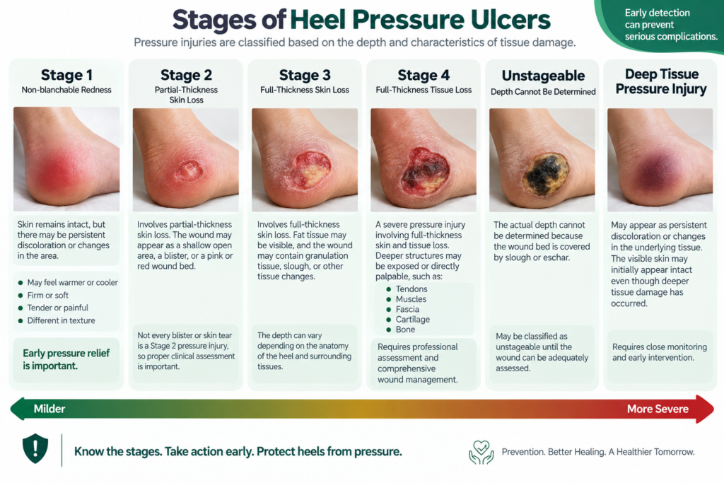
Visual guide to the different stages and classifications of heel pressure injuries.
Stage 1 Heel Pressure Ulcer
The skin remains intact, but there may be persistent discoloration or changes in the affected area.
The heel may also feel:
- Warmer or cooler
- Firm or soft
- Tender or painful
- Different in texture compared with surrounding skin
Early pressure relief is important at this stage.
Stage 2 Heel Pressure Ulcer
Stage 2 involves partial-thickness skin loss.
The wound may appear as:
- A shallow open area
- A blister
- A pink or red wound bed
Not every blister or skin tear is necessarily a Stage 2 pressure injury, so proper clinical assessment is important.
Stage 3 Heel Pressure Ulcer
Stage 3 involves full-thickness skin loss.
Fat tissue may be visible, and the wound may contain granulation tissue, slough, or other tissue changes.
The depth can vary depending on the anatomy of the heel and surrounding tissues.
Stage 4 Heel Pressure Ulcer
Stage 4 is a severe pressure injury involving full-thickness skin and tissue loss.
Deeper structures such as:
- Tendons
- Muscles
- Fascia
- Cartilage
- Bone
may become exposed or directly palpable.
These wounds require professional assessment and comprehensive wound management.
Unstageable Heel Pressure Injury
Sometimes the actual depth cannot be determined because the wound bed is covered by slough or eschar.
In these situations, the injury may be classified as unstageable until the wound can be adequately assessed.
Deep Tissue Pressure Injury
A deep tissue pressure injury may appear as persistent discoloration or changes in the underlying tissue.
The visible skin may initially appear intact even though deeper tissue damage has occurred.
How to Prevent Heel Pressure Ulcers
Prevention is especially important for people who spend long periods in bed or have limited mobility.
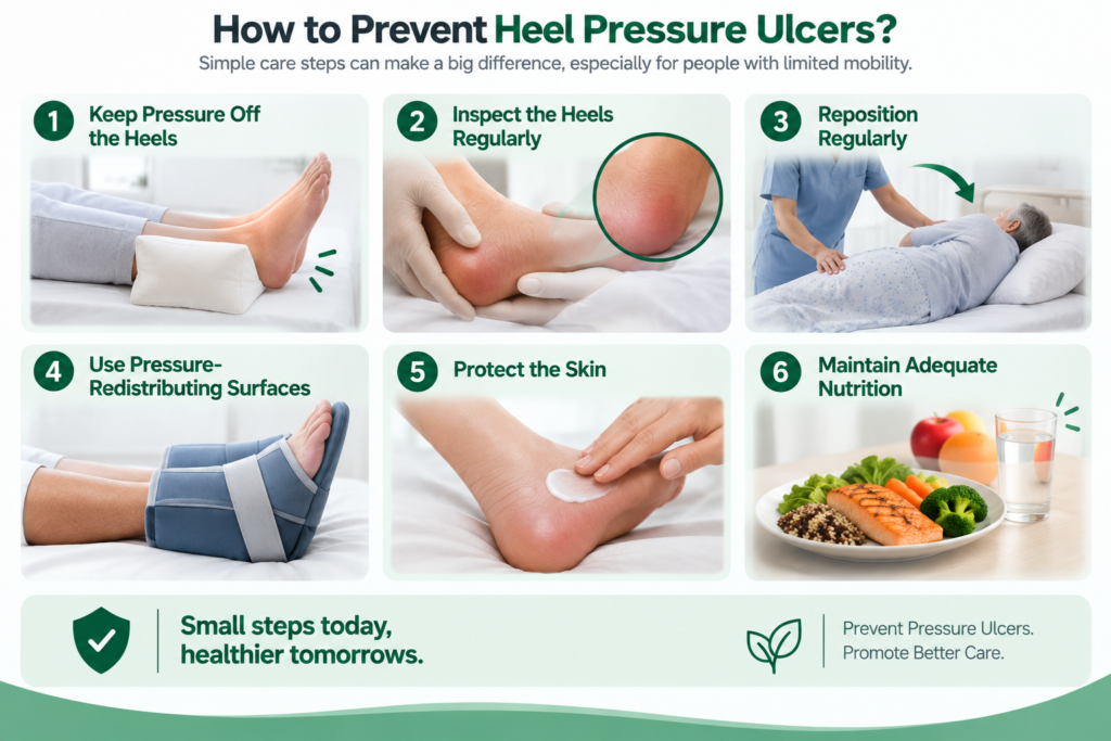
Key steps to help prevent heel pressure ulcers in people with limited mobility
1. Keep Pressure Off the Heels
One of the most important preventive strategies is heel offloading.
The heel should be completely relieved from contact pressure while the weight of the lower leg is supported along the calf. Specialized heel suspension devices or appropriate positioning methods may be used.
2. Inspect the Heels Regularly
Check the heels regularly for:
- Discoloration
- Swelling
- Blisters
- Skin breakdown
- Temperature changes
- Pain or tenderness
- Changes in texture
Regular inspection can help identify pressure damage before it becomes a deep wound.
3. Reposition Regularly
People with limited mobility should follow an individualized repositioning plan.
There is no single repositioning schedule that is suitable for everyone. The frequency should consider mobility, skin condition, support surface, medical condition, and individual tolerance.
4. Use Pressure-Redistributing Surfaces
Specialized mattresses, cushions, heel protectors, and support surfaces can help reduce or redistribute pressure.
However, these products should complement—not replace—regular repositioning and skin inspection.
5. Protect the Skin
Keep the skin clean and appropriately moisturized.
Manage excessive moisture promptly and avoid unnecessary friction when moving or repositioning the person.
6. Maintain Adequate Nutrition
Adequate nutrition and hydration support normal tissue maintenance and wound healing.
People at risk of pressure injuries may need nutritional assessment, particularly if they have poor appetite, unexplained weight loss, or difficulty meeting nutritional needs.
Heel Pressure Ulcer Care and Treatment
Treatment depends on the stage, wound depth, amount of drainage, tissue condition, infection risk, circulation, and the person’s overall health.
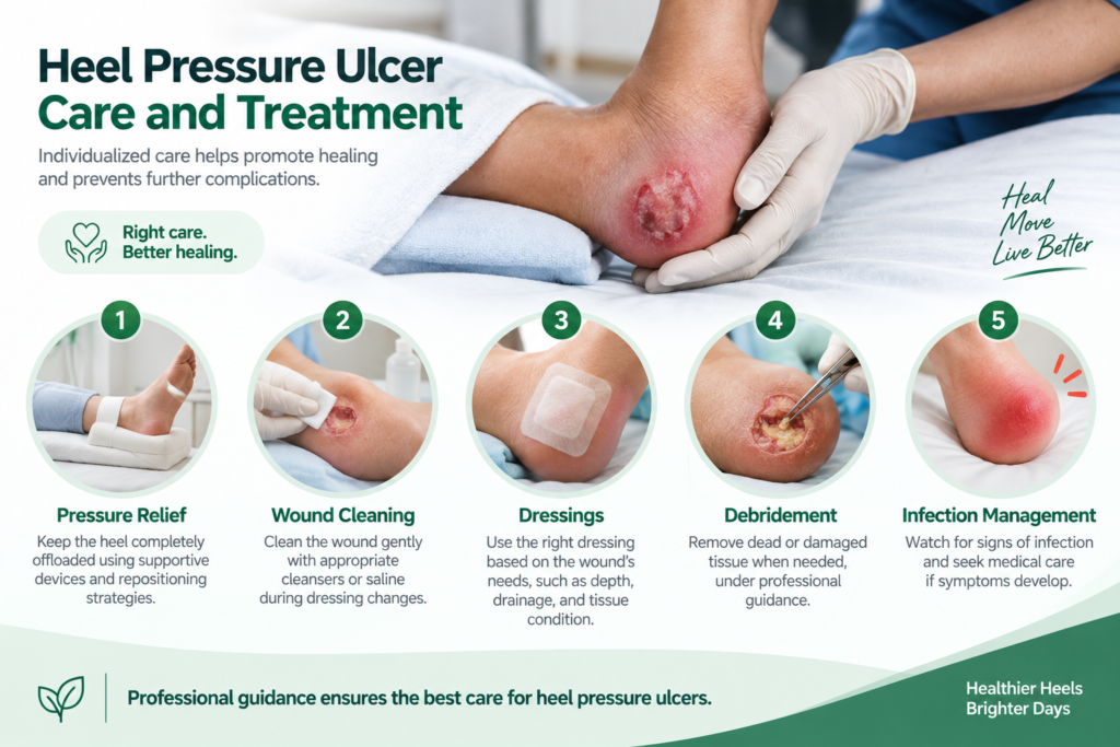
Key approaches used in the care and treatment of heel pressure ulcers.
Pressure Relief
Removing pressure from the affected heel is a fundamental part of care.
A healthcare professional may recommend:
- Heel suspension devices
- Pressure-redistributing mattresses
- Repositioning strategies
- Protective cushions
- Other offloading methods
Clinical guidance specifically recommends completely offloading vulnerable heels rather than allowing the heel to remain pressed against the bed surface.
Wound Cleaning
Open wounds may require appropriate cleansing during dressing changes.
Healthcare professionals may recommend wound cleansers or saline depending on the wound and clinical situation. Harsh substances should not be applied unless specifically recommended by a healthcare professional.
Dressings
The appropriate dressing depends on factors such as:
- Wound depth
- Amount of drainage
- Tissue condition
- Surrounding skin
- Infection risk
Different dressing options may be considered according to the wound’s individual needs.
Debridement
Some pressure ulcers contain dead or damaged tissue that can interfere with healing.
A trained healthcare professional may consider debridement when appropriate. However, heel wounds require careful assessment because blood supply and the presence of stable eschar can affect management decisions.
Infection Management
Pressure ulcers are not automatically infected, but infection can develop.
Possible warning signs include:
- Increasing redness
- Increasing warmth
- Swelling
- Worsening pain
- Pus-like drainage
- Foul odor
- Fever
- Rapid wound deterioration
Seek medical assessment if these symptoms develop.
How to Relieve Pressure on the Heel
Heel offloading is an important part of prevention and management.
A suitable heel suspension device can lift the heel away from the bed surface while supporting the lower leg. The device should be correctly fitted and should not create additional pressure on the Achilles tendon or behind the knee.
For some individuals, a properly positioned pillow or foam support may be used, but the exact method should be selected according to the person’s condition and clinical needs.
If a heel wound is already present, avoid putting direct weight or pressure on the affected area and seek professional guidance.
Can Heel Pressure Ulcers Heal?
Yes, heel pressure ulcers can heal, but healing time varies considerably.
Smaller and superficial injuries may improve when pressure is identified and relieved early. Deeper wounds can require prolonged treatment and specialized wound care.
Healing may also be affected by:
- Blood circulation
- Nutrition
- Infection
- Blood glucose control
- Mobility
- Wound depth
- Overall health
Some severe wounds may require advanced wound management or surgical treatment.
When Should You See a Doctor?
Professional assessment is recommended when a heel develops a new or worsening pressure injury.
Seek prompt medical attention if you notice:
- An open or deep wound
- Increasing redness or swelling
- Significant or worsening pain
- Pus-like drainage
- Foul odor
- Fever
- Black or severely discolored tissue
- Rapid wound progression
- Exposed tendon, muscle, or bone
Deep or non-healing heel wounds may require assessment by a wound-care specialist.
How Caregivers Can Help Prevent Heel Pressure Ulcers
Caregivers can play an important role in protecting vulnerable heels.
A practical routine may include:
- Inspect the heels regularly.
- Follow the person’s individualized repositioning plan.
- Keep the skin clean and appropriately moisturized.
- Manage moisture and incontinence promptly.
- Use recommended pressure-redistributing surfaces.
- Keep vulnerable heels appropriately offloaded.
- Encourage adequate nutrition and fluids when medically appropriate.
- Report new skin changes quickly.
- Follow the healthcare professional’s wound-care plan.
- Monitor for signs of infection or deterioration.
Heel protection should be individualized rather than relying on one routine for every patient.
A heel pressure ulcer is an area of skin and tissue damage caused primarily by prolonged pressure on the heel.
Early signs may include persistent discoloration, tenderness, swelling, or changes in skin temperature, firmness, softness, or texture.
Prevention includes regular skin inspection, individualized repositioning, heel offloading, pressure-redistributing surfaces, appropriate skin care, and adequate nutrition and hydration.
Yes. Increasing redness, warmth, swelling, pain, pus-like drainage, foul odor, fever, or rapid deterioration may indicate infection or another complication requiring medical assessment.
Healing time varies according to the stage, depth, circulation, nutrition, infection, pressure relief, and overall health. Deeper wounds generally require longer and more specialized care
Conclusion
Heel pressure ulcers can develop when prolonged pressure, friction, or shear damages the tissues around the heel. People with limited mobility, reduced sensation, poor circulation, diabetes, or prolonged bed rest may have a higher risk.
Early skin inspection, regular repositioning, and complete heel offloading are key parts of prevention. Once a pressure injury develops, treatment should be based on its stage, depth, circulation, infection risk, and the individual’s overall condition.
If a heel wound is open, worsening, deeply discolored, painful, draining, or showing signs of infection, professional medical assessment should be obtained promptly.
Medical Disclaimer: This article is for general educational purposes and does not replace diagnosis or treatment from a qualified healthcare professional.
Leave a Reply